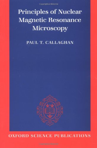
Principles of Nuclear Magnetic Resonance Microscopy epub
Par jones kevin le jeudi, octobre 27 2016, 07:22 - Lien permanent
Principles of Nuclear Magnetic Resonance Microscopy. Paul Callaghan

Principles.of.Nuclear.Magnetic.Resonance.Microscopy.pdf
ISBN: 0198539444,9780198539445 | 512 pages | 13 Mb

Principles of Nuclear Magnetic Resonance Microscopy Paul Callaghan
Publisher: Oxford University Press, USA
According to MR principles, the lowest detection limit is 1018 spins per voxel 24. We review recent efforts to detect small numbers of nuclear spins using magnetic resonance force microscopy. Force-detected nuclear magnetic resonance: recent advances and future challenges. UNIT V ELECTRO MAGNETIC RESONANCE AND MICROSCOPIC TECHNIQUES NMR – Basic principles – NMR spectrometer - Applications. Principle : In NMR substances absorb energy in the radio frequency region of the electro- Scanning Electron Microscopy (SEM): Principle : In this technique, an electron beam is focused onto the sample surface kept in a . Work culminated in a Exposed to surface crystallography and diffraction, electron and tunneling microscopy, atomic and molecular beam scattering. Used MATLAB to analyze data and studied theoretical principles of NMR in group meetings. Le Bihan studied NMR imaging intensively and suggested that diffusional movements of water and other molecules inside the body could be used to visualize microscopic structure and function of the organs and tissues being observed with MRI. This work, focusing on cell metabolism, led Damadian to believe that there should be a way to detect cancerous cells through chemical analysis rather than by relying on direct visualization under a microscope. The principles and measurement techniques underlying Diffusion MRI are rooted in the classic formula proposed for Nuclear Magnetic Resonance (NMR) by E. Ginseng cultivated in Hebei and Jilin of China, both of which were distinguished from extracts of P. Prior to the development of three-dimensional magnetic resonance imaging (MRI), the tracking of immune cell migration was limited to microscopic analyses or tissue biopsies. In addition, the similarity between 19F and 1H ( regarding their NMR properties) is a prerequisite for a meaningful overlay between the anatomical 1H MRI with the 19F MRI of labeled cells. �Nuclear magnetic resonance is a key technology for determining the structure of molecules and for visualising the anatomy of living tissue and microscopic structure. You are here: Home / Nanotechnology Books / Magnetic Microscopy of Nanostructures. NMR Imaging and Its Application to Clinical MedicineThe idea of using a magnetic gradient to introduce spatial information into signals from an NMR spectrometer, which can then be converted to an actu… Hébergé par OverBlog. This era saw a step change in his research productivity and impact, culminating in his first book, 'Principles of Nuclear Magnetic Resonance Microscopy' in 1994. H NMR metabolic profiling was performed to distinguish the water extracts of P. It has helped revolutionise “The NMR operates under the same principles as magnetic resonance imaging (MRI, which many patients experience as part of modern medical procedures) but is powered by a superconductor and has a magnetic field 400,000 times stronger than the Earth's magnetic field.